

Open ’til 7pm + Saturday
Call Us
832-804-7427
832-804-7427
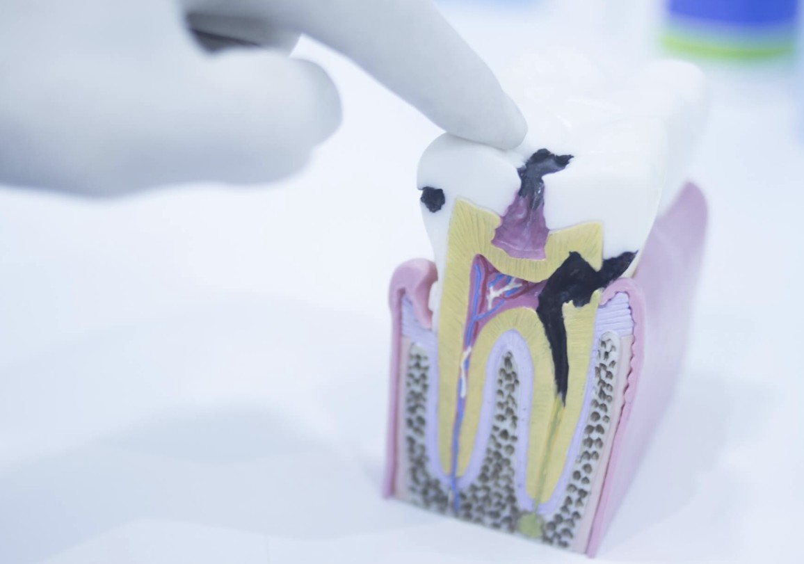
Cavities can look like chalky white spots, brown or black marks, pits, holes, or dark shadowing under the enamel. What you see depends on how early or advanced the decay is, where it is on the tooth, and whether the damage is on the surface or hidden between teeth.
A suspicious spot does not always mean a cavity. Stains, natural grooves, cracks, old dental work, and worn enamel can look similar, so a dentist may need an exam and X-rays to tell the difference.
Lovett Dental in Houston offers fillings and sealants and can provide the cavity-repair and protective services described here.
An early cavity often does not start as a black hole. It may first appear as a dull, chalky white area where the enamel has started losing minerals, even though the surface still looks intact.
These white spots often show up near the gumline, around braces, or in places where plaque collects often. In some cases, a fluoride treatment and other remineralization treatments may help strengthen early weakened enamel before a filling is needed, but only a dental evaluation can confirm that. For more on whether early decay can reverse, see Can a cavity heal?.
As decay progresses, a white area may turn tan, brown, or black. A dark spot is more concerning when it seems to be getting larger, sits in a rough or softened area, or appears with visible damage to the tooth.
Still, color alone is not enough to diagnose a cavity. Coffee, tea, tobacco, certain foods, and naturally deep grooves can stain healthy enamel, while some active cavities stay light-colored or are hard to see.
A more advanced cavity may look like a small pit, a rough opening, or a clear hole in the tooth. Food may catch there, and the edges may look white, brown, gray, or black.
When a cavity has formed a hole, the decay has usually moved beyond the outer enamel into deeper tooth structure. The visible opening may be smaller than the damage underneath, so the surface does not always show the full extent.
A white spot can be an early sign of mineral loss, especially if it looks matte or chalky instead of shiny. It can also come from enamel development differences, past fluoride exposure while teeth were forming, or harmless color variation.
A brown or black spot may be decay, but it may also be a stain. On back teeth, deep chewing grooves often hold pigment, so a dentist checks whether the area is still hard and intact or has started to break down.
A hole, pit, or chipped-looking area should be evaluated. It may be a cavity, a fracture, or worn enamel rather than decay alone.
Natural grooves are usually part of the tooth's normal shape and tend to look even and symmetrical. Cavities cause actual surface breakdown, not just a dark line or narrow crease.
Good home care, fluoride, and diet changes can help prevent tooth decay.
Cavities are easiest to notice on smooth front tooth surfaces and on chewing surfaces you can see clearly in a mirror. In good lighting, you may notice a white patch, a dark pit, or a visible opening.
Some areas are much harder to inspect at home. Decay can hide along the gumline, under plaque, around the edges of fillings, beneath crowns, or deep in the grooves of back teeth.
Cavities between teeth are often invisible without dental X-rays. A tooth can look normal from the front while decay is forming where two teeth touch.
A dentist may recommend X-rays when a cavity could be hidden between teeth or under the enamel. Bitewing radiographs are the standard method for finding interproximal decay, which often appears darker on the image because the damaged tooth structure is less dense.
Not every cavity needs an X-ray to be identified, and X-rays do not replace a dental exam. Dentists use them based on your age, cavity risk, symptoms, and what they see during the exam.
Decay can also form under or next to an existing filling or crown. In those cases, the restoration may block a direct view, which makes imaging especially helpful.
| Visible Change | What It May Look Like | How It May Differ from A Cavity |
| Surface stain | Flat yellow, brown, or black discoloration | Usually leaves the enamel hard and intact |
| Natural tooth groove | Thin dark line or narrow pit on a molar | May be normal anatomy without softened or missing enamel |
| Crack | Fine vertical, horizontal, or branching line | Usually looks more like a line than a rounded pit or hole |
| Worn enamel | Smooth, flattened, cupped, or translucent area | Often reflects gradual wear rather than a localized decay opening |
| Cavity | Chalky patch, discolored pit, shadow, or hole | May involve rough, softened, or missing tooth structure |
This comparison can help you know what to watch for, but it cannot confirm a diagnosis at home. More than one issue can affect the same tooth, and dentists may use lighting, magnification, instruments, and X-rays to tell them apart.
In children, early decay may appear as white, yellow, or brown areas near the gumline, especially on the upper front baby teeth. Cavities in back teeth may look like dark pits or openings in the chewing grooves, and they can spread faster because baby teeth have thinner protective layers.
In many cases, dental sealants for kids can help protect those grooves. This is one reason dentists watch dark or suspicious-looking grooves closely in children.
Adults may notice cavities around older fillings, along exposed root surfaces, or between teeth. Root cavities may look yellow, tan, brown, or black and often have less defined edges because roots are not covered by enamel.
At any age, a dark mark does not always mean advanced decay. A normal-looking tooth can still have a hidden cavity.

Schedule an exam if you notice a new white, brown, gray, or black area that does not go away, seems to grow, or appears with a pit or hole. A chipped-looking spot, food catching in the same place, or a change around a filling or crown also deserves attention.
Prompt care may help preserve more healthy tooth structure. Depending on what the dentist finds, you may need monitoring, preventive care for an early enamel change, fillings and sealants for a cavitated area, or another treatment if the damage is deeper. If a filling is needed, modern composite fillings are a tooth-colored option.
Seek urgent dental care if you have facial swelling, gum swelling with drainage, fever linked to a dental problem, or pain that is getting worse quickly. Trouble breathing or swallowing, swelling spreading into the face or neck, or severe illness needs immediate emergency care.
If a tooth change is persistent or unclear, do not rely on color alone or online photos. A professional exam can tell the difference between a stain, a normal groove, and decay, which is a good reason to visit the dentist.
Lovett Dental in Houston offers fillings and sealants and serves patients from nearby Baytown and Conroe; call (832) 804-7427 to schedule or book online.
Yes. Early mineral loss often looks chalky white, and some cavities stay light-colored for a time. A cavity does not have to be brown or black.
No. Black or brown spots can come from stains, deep grooves, certain foods or drinks, tobacco, or older dental work. A dentist can check whether the enamel is still hard and intact or has been damaged by decay.
Yes. Cavities can form between teeth, under the enamel, or around restorations without making an obvious hole. X-rays are often needed to find these hidden areas.
No. Probing a tooth at home can irritate the gums or damage a weakened area. It also cannot reliably tell you whether a spot is decay.
An early area of mineral loss may improve with professional guidance, fluoride, better plaque control, and fewer frequent sugar or acid exposures. Once the tooth has developed a true hole, it usually cannot rebuild itself and may need restorative treatment.
Lovett, Splendid & Haven Dentistry
Dental Offices in TX